
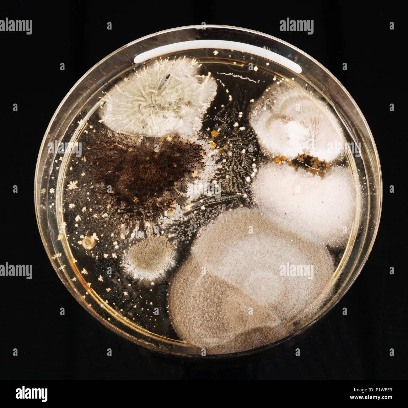
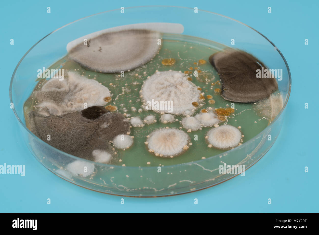
Characteristics and Different Shaped Colony of Bacteria and Mold Growing on Agar Plates . Stock
Hi guys! I could use Reddit's help in visually identifying the species of the mold growing on the three agar plates in the attached picture. The plates are Sabouraud's Dextrose Agar.
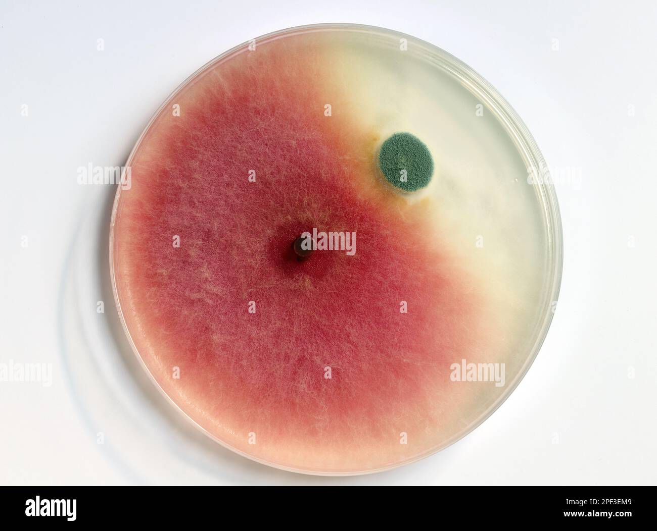
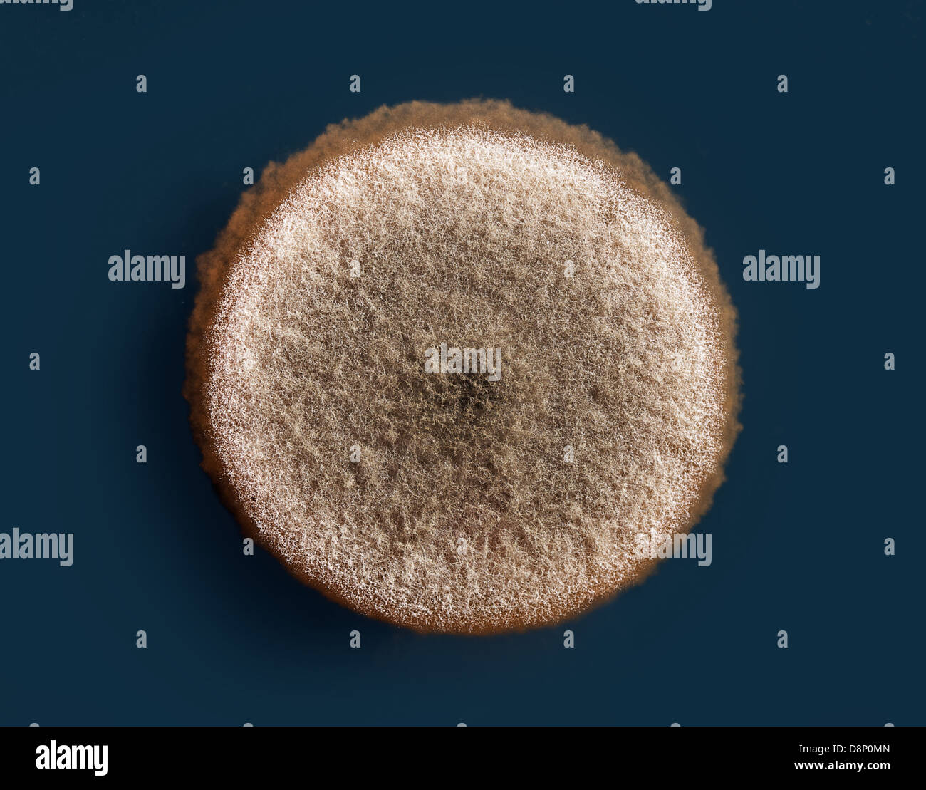
Culture of mold growing in a petri dish on a PDA, agar nutrient plate, colonies of Fusarium (red
Ready-to-use Potato Dextrose Agar Plates are a general purpose medium for the cultivation of fungi, including yeasts and molds. It is used for pharmaceutical product sterility tests according to the United States Pharmacopeia (USP) and for plate count enumeration tests for dairy products, foods, and…. Compare this item.
growth of in a Petri dish, Bacteria, yeast and mold growing on an agar plate
Step 1: Prepare Your Agar Plates. Agar is a seaweed based gelatin that is used as the medium to grow microbes. Start by mixing 1 tablespoon of agar with one cup of COLD water in a small pot. (If the agar is mixed into the water after it is heated, the agar will clump together unevenly.)
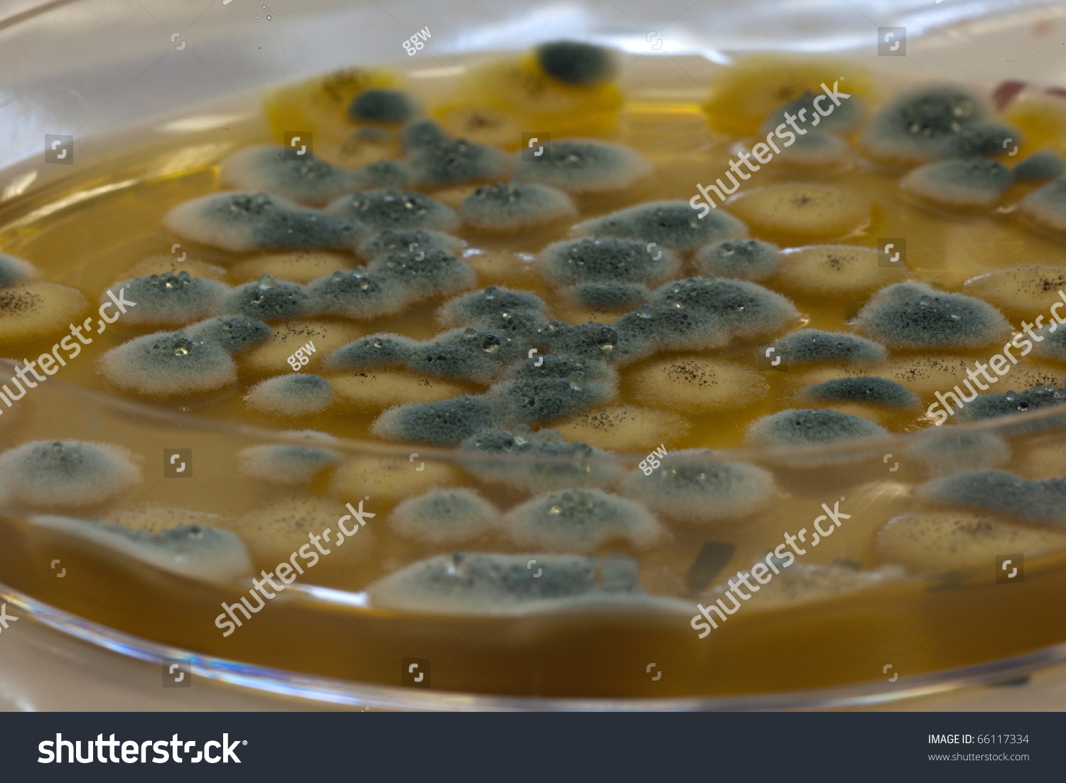
Macro Mold Growing On Agar Plate Stock Photo 66117334 Shutterstock
Colony morphology is a method that scientists use to describe the characteristics of an individual colony of fungi growing on agar in a Petri dish. It can be used to help to identify them. Colony morphology. A circular piece of bread that has been allowed to go mouldy. Plate 1 contains a circular piece of bread that has been allowed to go mouldy.

Macro Mold Colonies Growing On An Agar Plates. Stock Photo Image 20410960
Remove lid from test plate and set aside. Gently tap the item you wish to test with the plate 1-2 times. Make sure that the agar material is exposed to the item being tested when tapping. The puffs of air created from the tapping motion will cause any mold spores embedded in the item to attach to the agar material. Place lid back on test plate.
Macro Mold Colonies Growing On An Agar Plates. Stock Photo Image of biotechnology, antibiotic
The 3M™ Petrifilm® Rapid Yeast and Mold Count Plate method is faster and provides results in as little as 48 hours of incubation time, vs. the traditional agar method that requires a five day incubation period. This plate features a new indicator technology that makes colonies easier to interpret.
growth of in a Petri dish, Bacteria, yeast and mold growing on an agar plate
mold's natural defense mechanism against other molds and microorganisms, but,. Compare the mold growth from your kit with the images below to help identify the mold on your petri plates. Remember that a laboratory test is the most accurate method for mold. On agar petri dishes, it usually appears cream to pink colored initially, but then.

Characteristics and Different Shaped Colony of Bacteria and Mold Growing on Agar Plates . Stock
Petri dishes (also called settling plates) are designed to grow mold spores and since there are mold spores in the air everywhere all the time, you should expect to get mold spores growing on the plates. But since plates can only capture a limited group of spores (i.e., the ones closest to the plate and the heaviest ones that are more likely to.
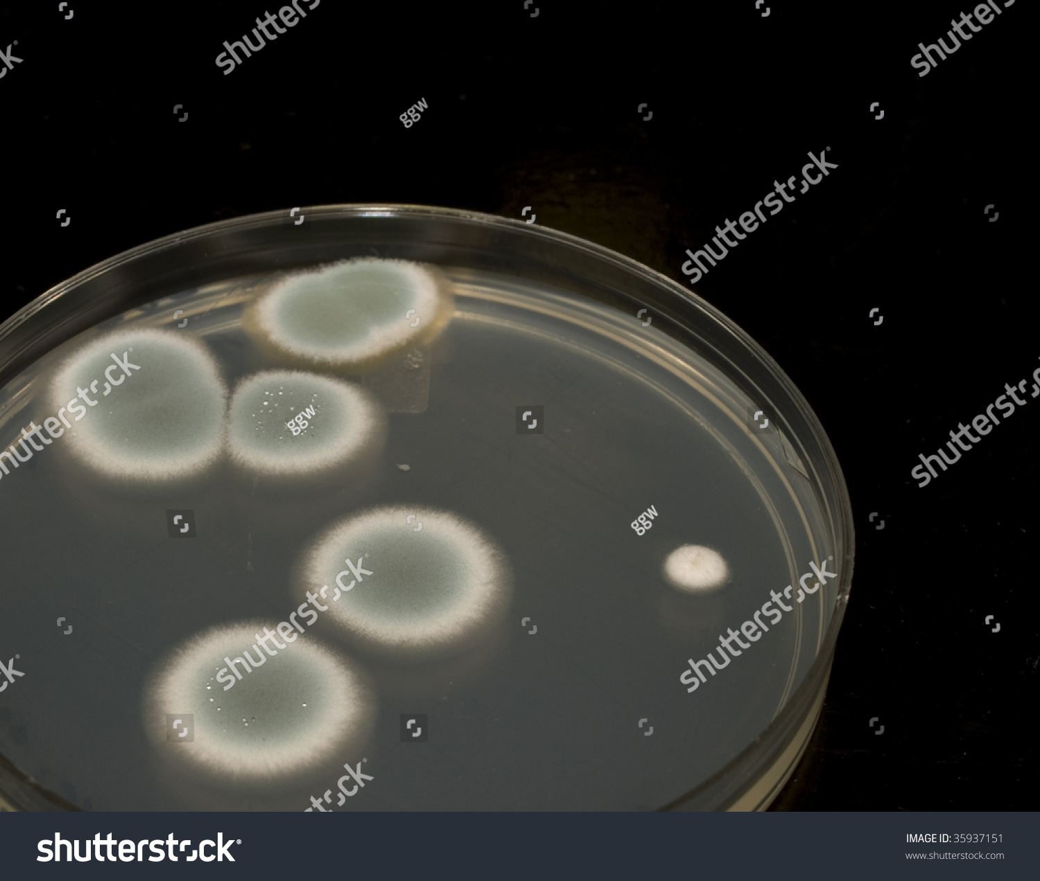
Mold Colonies Growing On An Agar Plate. Stock Photo 35937151 Shutterstock
Wash & dry your hands thoroughly prior to testing. Hold the plate so the yellow agar is facing towards the object or pet. Tap the object in 4 different representative places (The yellow agar should not touch the object). This will force the mold spores into the air and onto the plate. How-To Sample: Visible Mold.
Interesting mold contamination on my agar plate MoldlyInteresting
Place loop or wire material portion of "fungus" colony onto slide with a drop of stain, and add a coverslip. Gently press down to flatten potential mold and agar. Use a paper towel dipped into alcohol for pressing. Discard towel and wipe hands with disinfectant and dry.
Mold Colonies Growing On An Agar Plates. Stock Photo 36073933 Shutterstock
Question: shook mold off clothing onto a culture plate - Candida, possibly Candida auris? I tapped an agar plate (MEA w/chloramphenicol) against some contaminated clothing. It grew on the plate and on the scotch tape adhesive around the plate. (a milky look) - Photos attached.

Characteristics and Different Shaped Colony of Bacteria and Mold Growing on Agar Plates from
Yeast and Mold Count Plates identify food borne yeast and mold in your food products and in your food processing environment. Yeasts are easily differentiated from molds on the plate: - Yeasts are typically indicated by small, blue-green colonies with defined edges and no foci. - Molds are…. Compare this item.

Characteristics and Different Shaped Colony of Bacteria and Mold Growing on Agar Plates from
Yeast and Mold Agar should be sterilized without pH adjustment and sterile acid added to the medium cooled to 45 - 50°C. Acidified Yeast and Mold Agar should not be heated.. Plates per 500 g: 670.00 Preparation: Suspend 41 grams of the medium in one liter of purified water. Heat with frequent agitation and boil for one minute to completely.

Green Mold Agar Plate Stock Photo 417683947 Shutterstock
Wash & dry your hands thoroughly prior to testing. Hold the plate so the yellow agar is facing towards the object or pet. Tap the object in 4 different representative places (The yellow agar should not touch the object). This will force the mold spores into the air and onto the plate. How-To Sample: Visible Mold.

Mildew Culture On Agar Plate Laboratory Scene HighRes Stock Photo Getty Images
Method for Yeasts and Molds: Agar: Although many selective agars exist for the cultivation and determination of mold and yeast cultures, a majority of them do not depend on strict nutritive requirements for growth. Many fungal strains will grow on Sabouraud Dextrose Emmons Agar. Alternative agars for growth include Sheep Blood Agar, Nutrient.
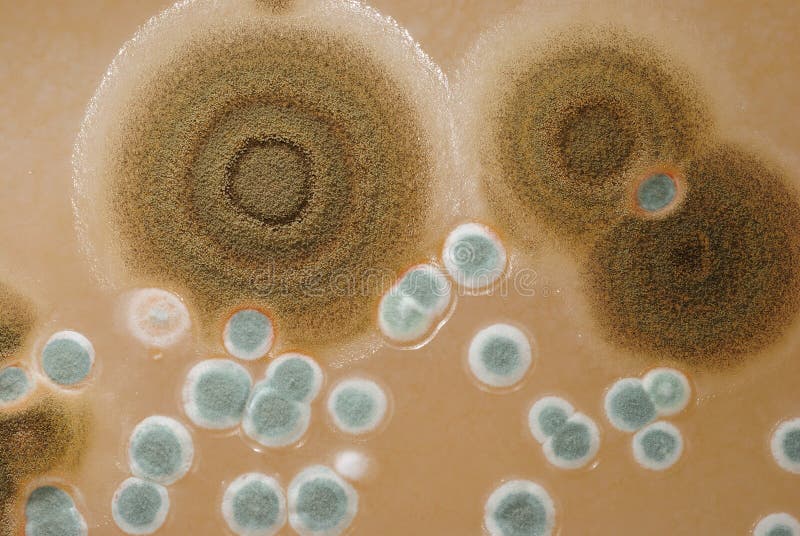
a mold colony on an agar plate (aspergillus niger Stock Photo Alamy
What Can Grow on a Nutrient Agar Plate? Bacteria. Each distinct circular colony should represent an individual bacterial cell or group that has divided repeatedly. Being kept in one place, the resulting cells have accumulated to form a visible patch. Most bacterial colonies appear white, cream, or yellow in color, and fairly circular in shape.
